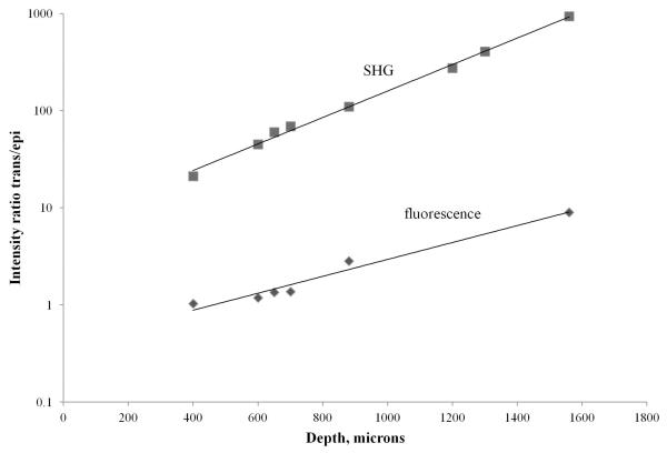
Figure 5.
Plot of the intensities ratio for SHG and fluorescence images acquired by the trans-PMT and epi-PMT as a function of imaging depth. The images were acquired from a collagen matrix embedded with MDA-MB-231cells expressing paxillin-GFP. The collagen matrix was extracted from the well and placed in a standard 35 mm Petri-dish with a glass window. The thickness of the sample was about 4 mm.

