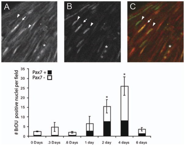
Figure 2.
Pax-7 expression and BrDU incorporation during androgen treatment of newly metamorphosed animals. (A) After 2 days of androgen treatment, BrDU has accumulated in nuclei shown in confocal image taken parallel to muscle fibers of whole mount larynges. Nuclei are elongated and are similar to the morphology of Pax-7 immunoreactivity (B) in the same location. Pax-7 and BrDU immunoreactivity overlap (A–C, arrow), but not all BrDU positive cells are Pax-7 positive (A–C, arrowhead), and not all Pax-7 cells are BrDU positive as is shown in an overlay of Pax-7 (red) and BrDU (green) labeling (C). (D) Counts of cells show that the number of BrDU positive cells increases during androgen treatment (error bars show SEM, * denotes statistically significant difference from day 0 of p < 0.05 for Student’s t-test after Bonferroni correction for multiple comparisons). Throughout androgen treatment, BrDU accumulating cells include cells that are Pax-7 positive, as well as those that are Pax-7 negative (black bars show proportion of BrDU positive cells that were also positive for Pax-7.

