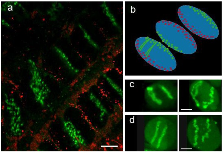
Figure 2.
Rabl chromosome organization in wheat root tissue. (a) Centromeres (green) and telomeres (red) are labelled by fluorescence in situ hybridization (FISH) and are located at opposite sides of the nuclei; (b) Diagrammatic interpretation of the organization in (a); (c) Introgressed pair of rye chromosomes in wheat labelled by genomic in situ hybridization (GISH) with total rye genomic probe confirms a Rabl organization of individual CTs. (d) Single rye arm translocation into wheat localized by GISH. In (c) and (d) the RH panel in each case shows a nucleus from a seedling treated with 5-AC, whereas the LH panel shows a control untreated seedling. The 5-AC treatment has disrupted the CT organization, but the CTs remain in a Rabl configuration. Bar = 10 µm.

