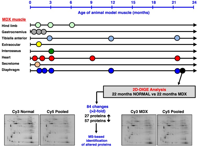
Figure 2.
Mass spectrometry-based proteomic profiling of the mdx mouse model of Duchenne muscular dystrophy: The upper panel of the figure summarizes the types of muscle and age groups of mdx mice that have been used over the last few years to identify global changes in the dystrophic muscle proteome. All listed proteomic studies have been recently discussed in comprehensive reviews of the proteomics of the dystrophin-glycoprotein complex and dystrophinopathy [50,69]. The lower panel gives an overview of the difference in-gel electrophoretic (DIGE) analysis of the aged mdx diaphragm muscle presented in this report.

