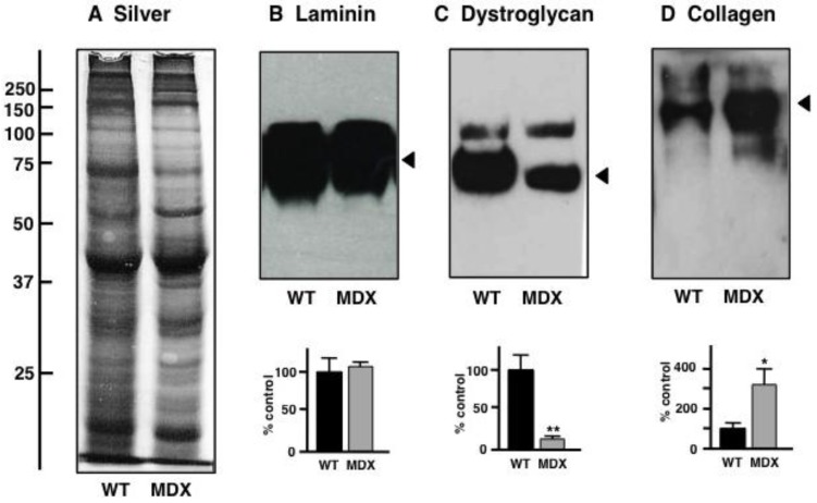
Figure 7.
Gel electrophoretic and immunoblot analysis of the aged mdx diaphragm muscle: Shown is a silver-stained gel (A) and representative immunoblots (B–D) with expanded views of antibody-decorated bands. Lanes 1 and 2 represent preparations from non-dystrophic wild type (WT) versus dystrophic (MDX) diaphragm muscle, respectively. Immuno-decoration was carried out with primary antibodies to laminin (B), the dystrophin-associated glycoprotein β-dystroglycan (C) and collagen (D). Below the individual immunoblots are shown panels, which give a graphical representation of the immuno-decoration levels in normal versus mdx preparations (Student’s t-test, unpaired; n = 4; * p < 0.05; ** p < 0.01).

