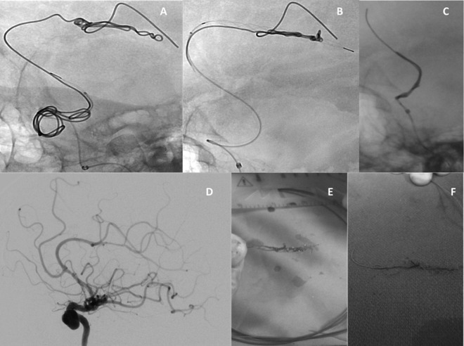
Figure 1.
Digital subtraction angiography showing capture of (A) the proximal segment and (B) the distal segment in the coil and (C) retrieval of the coil (Penumbra Complex Standard) by retracting the stentriever (Trevo ProVue) back into the guide catheter with the coil ensnared. (D) Postoperative imaging shows mild vasospasm (arrow) and no evidence of vascular injury or branch occlusion. (E, F) The interwoven coil and Trevo ProVue after retrieval.

