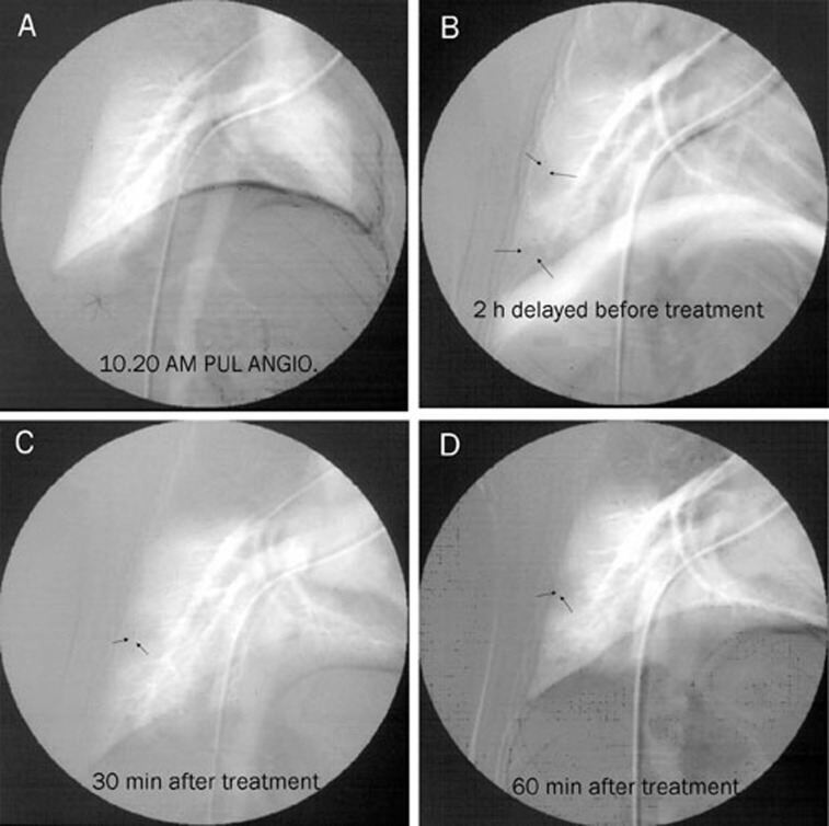
Figure 3.
Digital subtraction angiography in APT. Arrows indicate thrombi in the right pulmonary artery. (A) Before pulmonary thromboembolism; (B) Two hours after pulmonary thromboembolism. The right pulmonary artery shows a complete obstruction. (C) FIIa treatment for 30 min. A reduction in obstruction was detected in the lower lobar pulmonary artery branches. (D) FIIa treatment for 60 min. The pulmonary emboli almost completely disappeared.

