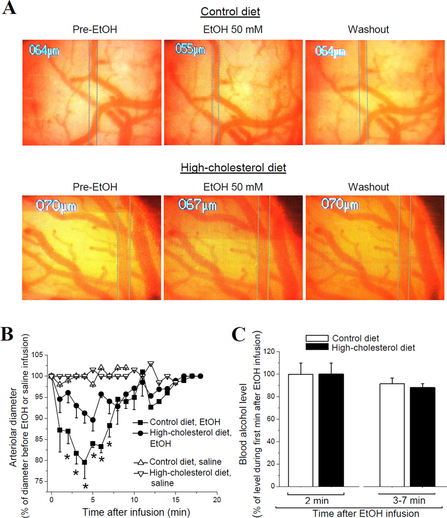
Figure 1.
Dietary cholesterol protects against alcohol-induced pial arteriole in vivo constriction independently of EtOH elimination rate from the systemic compartment. (A) Screenshots of brain surface obtained through a closed cranial window. Vertical dashed lines highlight external diameter of pial arteriole. Pial arteriole diameter was monitored before (left) and after (middle) a 50 mM EtOH infusion into cerebral circulation via catheter in carotid artery. Washout of EtOH was achieved by infusion of sodium saline (right). (B) Averaged data show a significant decrease in pial arteriole diameter (PAD) in response to EtOH in rats on high-cholesterol diet (n=4) compared to control group (n=4). *p<0.05, when compared to EtOH-induced arteriole constriction in control group. (C) Averaged blood alcohol levels after infusion of 50 mM EtOH into cerebral circulation of rats on control vs. high-cholesterol diet. Data show lack of significant differences between the groups throughout 7 min following EtOH infusion. Each data point was obtained from ≥4 animals.

