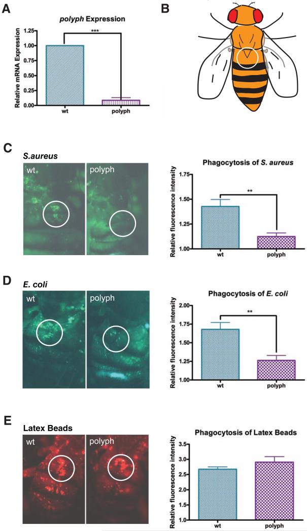
Figure 1. polyph, a putative amino acid transporter, is required for microbial phagocytosis.
(A) Comparison of polyph transcript levels via qPCR in wildtype flies (wt) and flies containing a transposon insertion in the polyph gene (polyph). Relative expression was measured using rp49 as an endogenous control. A pool of ten flies per genotype was used in each experiment. (B) Representation of how the fly is visualized during the in vivo adult phagocytosis assay. The encircled area represents the area of the dorsal vein around which the sessile blood cells congregate. (C, D, E) Representative pictures depicting phagocytosis in wt and polyph flies of (C) fluorescein-labeled S.aureus bioparticles, (D) fluorescein-labeled E.coli bioparticles, and (E) red fluorescently labeled latex beads. Approximately 6 flies per genotype were used in each experiment. Quantification follows. Error bars, ±SE. ** p<0.01, *** p<0.001.

