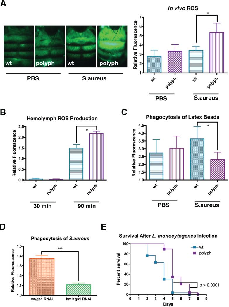
Figure 3. polyph has increased ROS in its hemolymph and decreased phagocytosis when exposed to bacteria.
(A) Representative pictures of oxidized CM-H2CDFDA-derived fluorescence in wt and polyph flies after a 30 minute preinjection of either PBS or overnight culture of S.aureus. Approximately 6 flies were used per genotype in each experiment. Quantification follows. (B) Measurement of oxidized CM-H2CDFDA-derived fluorescence in wt and polyph blood cells incubated ex vivo with an overnight S.aureus culture. (C) Quantification of the phagocytosis of red fluorescently labeled latex beads in wt and polyph flies after a 30 minute preinjection of either PBS or overnight culture of S.aureus. Approximately 6 flies per genotype were used in each experiment. (D) Quantification of the phagocytosis of fluorescein-labeled S.aureus bioparticles in wt/gs1 RNAi and hml>gs1 RNAi. Approximately 6 flies per genotypewere used in each experiment. (E) Representative survival curve of wt and polyph flies after injection of L.monocytogenes (OD 0.1). n = 28-30 flies All experiments were done in triplicate. Error bars, ±SE. *p<0.05.

