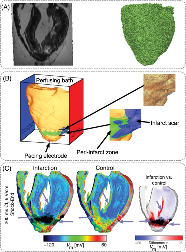
Figure 5.

MRI-based model of healed infarction in the rabbit. (A) Ex-vivo MRI scan of the rabbit heart with healed myocardial infarction and anterior view of the fibre orientations in the ventricles. (B) Left panel, anterior view of the ventricles submerged in a perfusing bath and placed between plate electrodes (blue, grounding electrode; red, shock electrode). The infarct scar is shown in blue, the peri-infarct (border) zone is shown in green. The pink square at the apex shows the location of the pacing electrode. The bottom inset shows the highly detailed structure of the scar and the PZ. The top inset shows the details of the computational mesh. (C) Distribution of shock-end Vm. Less tissue was excited in the infarction model (purple arrows). Right-most panel shows the Vm difference between infarction and control models, computed as control Vm minus infarction Vm. Images based on figures published in Rantner et al.6
