
Figure 4.
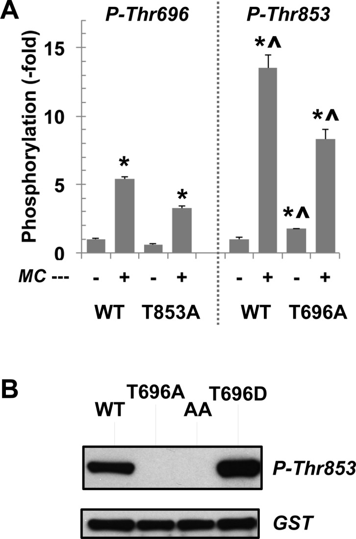
Phosphorylation of MYPT1 by ROCK. (A) WT, T696A, and T853A MLCPs were phosphorylated for 30 min at 30 °C with ROCK (36 milliunits) in the absence or presence of 1 μM MC-LR and subjected to immuno-dot blotting using antibodies for total, P-T696, and P-T853 MYPT1. Potential contamination of PP2A was eliminated with OA (10 nM) in the mixture. The mean value ± SEM of staining density of phospho-Thr696 or Thr853 vs total MYPT1 was obtained from triplicate assays. An asterisk and a carrot indicate p < 0.05 against the value without MCLR and the value of P-Thr696, respectively. (B) GST-tagged MYPT1 fragment (residues 654–880) phosphorylated for 30 min at 30 °C with ROCK (36 milliunits) and subjected to immunoblotting. Representative data from duplicate assays are shown.