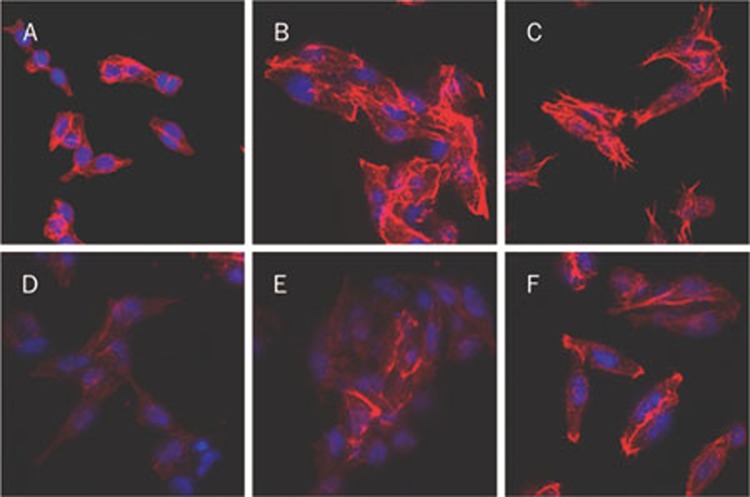
Figure 2.
Fluorescence photomicrographs of SW620 cells with rhodamin-conjugated phalloidin and Hoechst 33342 staining (×400). SW620 cells were placed in 24-well culture dishes at 3×10⁴ cells/well and treated with different conditions for 1 h. Then, the cells were fixed with 4% paraformaldehyde and washed with PBS three times. The DNA was stained with Hoechst 33342 (blue) and the actin cytoskeleton with rhodamin-conjugated phalloidin (red) which were observed under a laser scanning confocal fluorescence microscope. (A) Media only; (B) With 100 μmol/L of PAR2-AP; (C) With 10 nmol/L of factor VIIa; (D) With 100 μg/mL of EGCG; (E) With EGCG (100 μg/mL)/PAR2-AP (100 μmol/L); (F) With EGCG (100 μg/mL)/factor VIIa (10 nmol/L).

