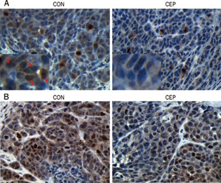
Figure 6.
Immunohistochemical analyses of STAT3. After treatment of CEP for 20 d, the tumor samples were taken from the sacrificed mice and immunostained with antibodies against total and phosphorylated STAT3. (A) Immunohistochemical staining of total STAT3. STAT3 nuclear localization in control group was indicated with the red arrows. (B) Immunohistochemical staining of phospho-STAT3. Original magnification: ×400.

