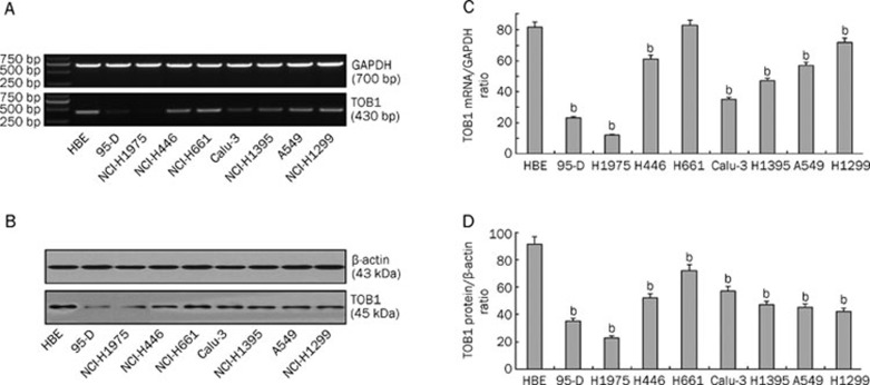
Figure 1.
TOB1 expression is variably decreased in eight lung cancer cell lines. (A) Expression of TOB1 mRNA in lung cancer cell lines assayed via RT-PCR; GAPDH was used as the loading control. Total RNA was extracted from normal and lung cancer cells. The 430 bp TOB1 cDNA fragments were separated and visualized by 1% agarose gel electrophoresis and ethidium bromide staining. (B) Expression of TOB1 protein in normal epithelial cell line HBE and eight lung cancer cell lines, separately assayed via Western blot analysis with β-actin as the loading control. Whole cell lysates (50 μg) from each of the nine cell lines were separated using SDS-PAGE and transferred onto PVDF membranes. Bands were visualized using monoclonal anti-TOB1 antibodies with a chemiluminescence detection system. All experiments were performed independently at least three times. Mean±SD. bP<0.05 vs HBE cells.

