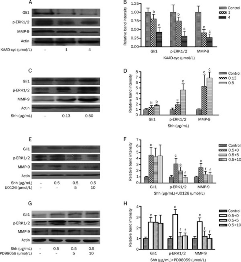
Figure 5.
Effects of KAAD-cyc, Shh, U0126, or PD98059 on the expressions of Gli1, p-ERK1/2, and MMP-9 proteins in Bel-7402 cells. (A) Western blot analysis of Gli1, p-ERK1/2, and MMP-9 protein levels in cell lysates from Bel-7402 cells treated with 1 or 4 μmol/L KAAD-cyc for 24 h. (B) The values under each lane indicate relative density of the band in Figure 5A normalized to β-actin, respectively. (C) Western blot analysis of Gli1, p-ERK1/2, and MMP-9 protein levels in cell lysates from Bel-7402 cells treated with 0.13 or 0.50 μg/mL Shh for 24 h. (D) The values under each lane indicate relative density of the band in Figure 5C normalized to β-actin, respectively. (E) Western blot analysis of Gli1, p-ERK1/2, and MMP-9 protein levels in cell lysates from Bel-7402 cells pretreated by 0.50 μg/mL Shh, then treated with 5 or 10 μmol/L U0126 for 24 h. (F) The values under each lane indicate relative density of the band in Figure 5E normalized to β-actin, respectively. (G) Western blot analysis of Gli1, p-ERK1/2, and MMP-9 protein levels in cell lysates from Bel-7402 cells pretreated by 0.50 μg/mL Shh, then treated with 5 or 10 μmol/L PD98059 for 24 h. (H) The values under each lane indicate relative density of the band in Figure 5G normalized to β-actin, respectively. bP<0.05, cP<0.01 vs control; eP<0.05, fP<0.01 vs Shh.

