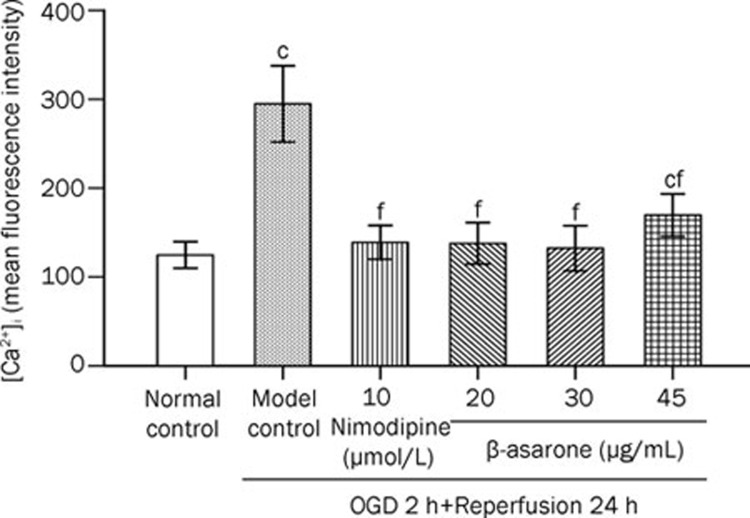
Figure 3.
Effect of β-asarone on [Ca2+]i in OGD/R treated PC12 cells. Model control cells were treated with 2 h OGD followed by 24 h reperfusion. The treated cells were incubated with nimodipine (10 μmol/L) or β-asarone (20, 30, or 45 μg/mL) 1 h before OGD and 2 h throughout OGD, followed by their transfer to full and drug-free culture medium under normoxic conditions for 24 h. Normal control cells were incubated in a regular cell culture incubator under normoxic conditions. After these treatments, [Ca2+]i was analyzed using flow cytometry. The mean fluorescent intensity of Fluo-3 was used as the indication of [Ca2+]i quantity. Mean±SD for 10 samples. cP<0.01 vs normal control group. fP<0.01 vs model control group.

