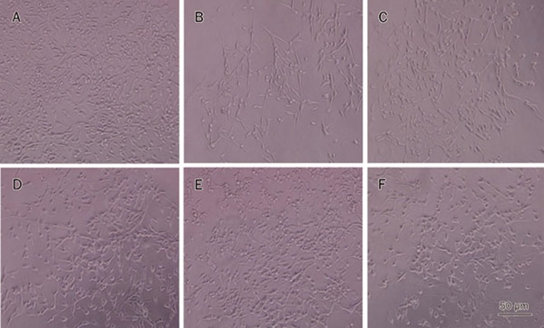
Figure 4.
Cellular morphology was observed under inverted phase contrast microscope. Normal neuronal morphology was seen in the bright field images (A, ×200). The number of cells showed a significant reduction. The cells exhibited round, slender and degenerated morphology after OGD/R (B, ×200). The number of cells showed a dramatic increase. The cells exhibited improved cellular morphology after treatment with β-asarone (20, 30, or 45 μg/mL) (D, E, F, ×200) or nimodipine (10 μmol/L) (C, ×200).

