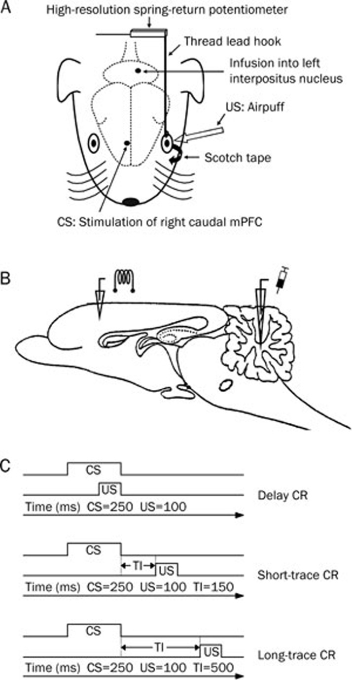
Figure 1.
Experimental design. (A) The upper left eyelid movements were measured by a high-resolution spring-return potentiometer that was attached via a thread lead that was hooked through a nylon loop, which was sutured into the left upper eyelid, and the left lower eyelid was taped open. One electrode was implanted in the right caudal medial prefrontal cortex (mPFC) and one infusion guiding cannula was implanted in left cerebellar interpositus nucleus. Electrical stimulation of right caudal mPFC was used as a conditioned stimulus (CS), and airpuff was presented to the left cornea as an unconditioned stimulus (US). (B) Diagram of the sagittal section of guinea pig brain, showing the stimulating and infusion sites. (C) Schematic diagram showing the delay, short-trace, and long-trace eyeblink conditioned response (CR) paradigms used in the present study. Note that the CS, US, and total trial lengths were equal in each conditioning. Short and long-trace CR were different in trace interval (TI) length.

