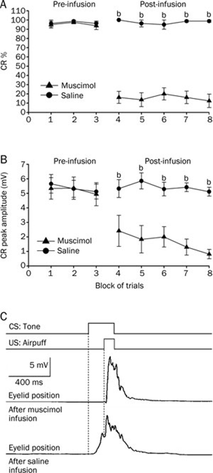
Figure 4.
Delay conditioned group data for the effects of muscimol (▴) and saline (•) infused into the left cerebellar interpositus nucleus. (A) Infusion of muscimol abolished the responses almost completely as illustrated by its effects on the percentage of trials in which the delay CRs were seen, whereas infusion of saline had no significant effect on the delay CRs. (B) Muscimol infusion significantly decreased the peak amplitude of the delay CRs. (C) Eyelid position of an animal after muscimol and saline infusion in the sixth trial. Upper panel: the conditioning paradigm illustrating the timing of the CS and the US. Middle panel: eyelid position after muscimol infusion. Lower panel: eyelid postion after saline infusion. All data are from the same animals. Mean±SEM. n=8. bP<0.05 vs control.

