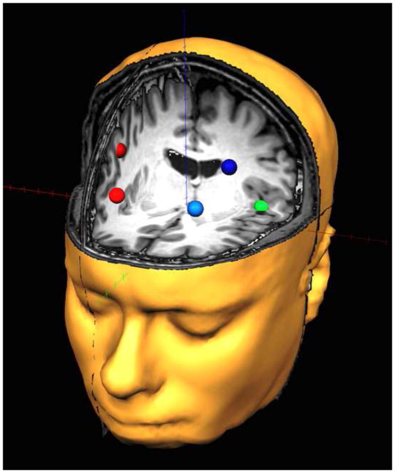
Fig. 1.

Dorsolateral prefrontal and auditory regions of interest. The red (right hemisphere) and blue (left hemisphere) nodes represent anterior and posterior regions of the prefrontal cortices corresponding to the regions that were focused upon. The green sources represent the auditory cortical regions and corresponded to the anatomical regions of heschl’s gyri (right auditory cortex is not shown). Note that the time series of each node reflects the average neuronal activity over that brain region, and not the amount of activation at a precise neuroanatomical coordinate (e.g., a voxel in Montreal Neurological Institute space).
