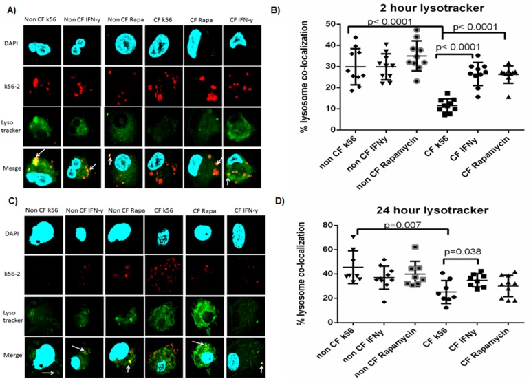
Figure 6. Lysosomal co-localization is increased with IFN-γ in CF.
6A) Confocal microscopy for non-CF and CF macrophages infected with k56-2. Macrophages were labeled with green lysotracker and bacterial co-localization to lysosomes was measured after a 2 hour infection. Macrophage nuclei are stained blue with DAPI. Co-localization of bacteria with lysotracker is noted in yellow by white arrows. 6B) Quantitative scoring of % bacterial co-localization with lysosomes from 6A, n = 8 subjects, Mann-Whitney testing. 6C) 24 hour k56-2 infection with lysotracker staining. 6D) Bacterial co-localization with lysotracker quantitative scoring for 5C, n = 8 subjects, Mann-Whitney testing.

