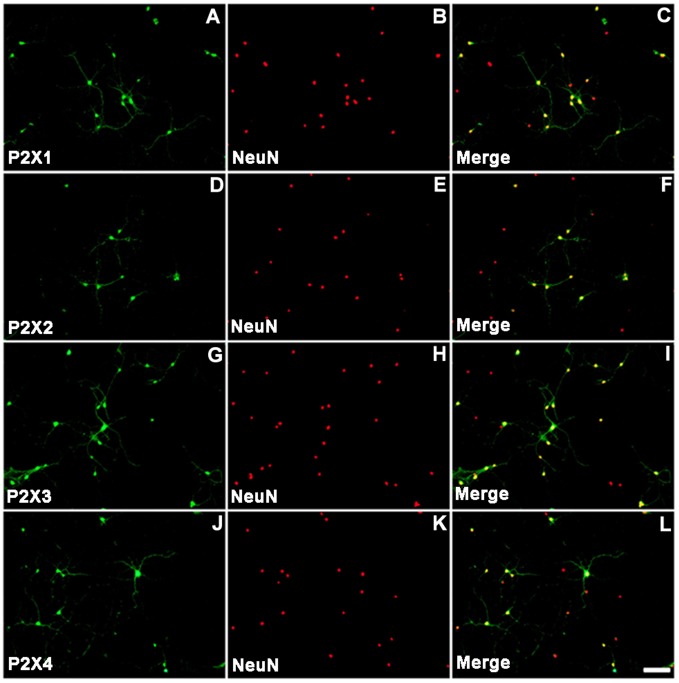
Figure 4. Immunofluorescent staining of cultured NG neurons.
NG neurons were stained using antibodies against P2X1, P2X2, P2X3, and P2X4 receptor subunits (A, D, G, and J, respectively). The nuclei of cultured NG neurons were stained with antibodies against NeuN (B, E, H, K). Merged images (C, F, I, L) representing co-staining of P2X receptor subunits and NeuN are shown. The scale bar shown in L is representative of all images, and represents 50 µm.

