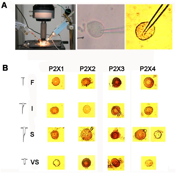
Figure 8. Relevance of the P2X1–4 subunits on the four types of IATP.

(A) Schematic view of the setup for the whole cell patch clamp and a representative image of a recorded cell under the phase contrast microscope and immunohistochemistry. (B) Immunohistochemistry revealed positive or negative staining for P2X1–4 subunits, which correlated with the type of IATP and cell size. The samples in each row were from four different neurons that responded to ATP with different types of ATP-activated current. P2X3 staining was positive in all four types of IATP neurons. P2X1 was positive in F, I, and S IATPs, but negative in VS. P2X2 staining was only absent in neurons with type I IATP, and P2X4 was positive in neurons with type F, I, and some S IATPs.
