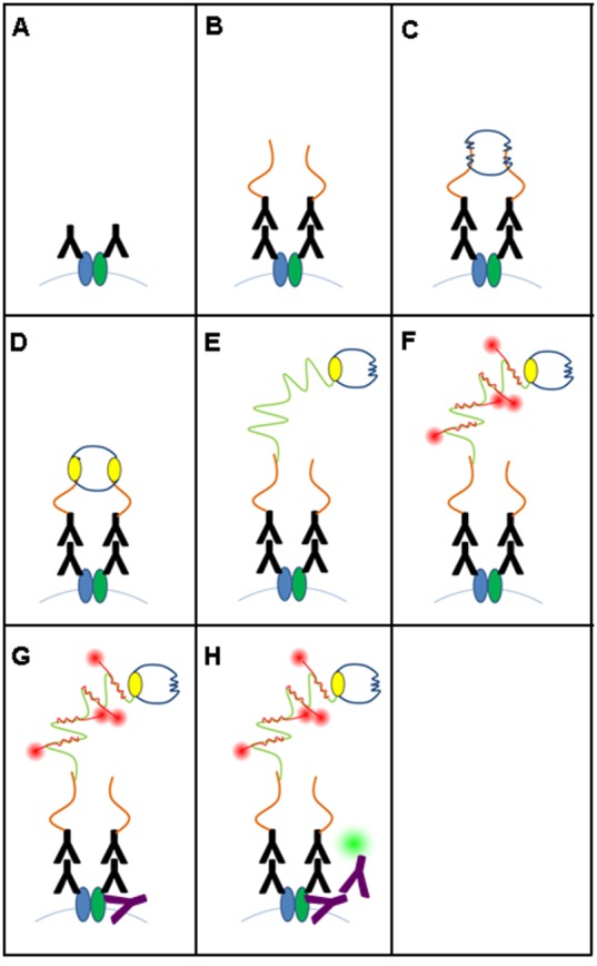
Figure 1. Schematic drawing of different antibody binding and visualization steps of isPLA combined with IF.

(A) Primary ABs of the isPLA bind to two different proteins, which are expected to colocalize. (B) Secondary ABs conjugated with oligonucleotides bind to the primary ABs (PLA Probe PLUS and PLA Probe MINUS). (C) Two more oligonucleotides (blue) hybridize to the two PLA probes. (D) Ligase (yellow) joins the two added oligonucleotides to form a closed circle. (E) Polymerase (yellow) induces a rolling circle amplification (RCA) using the ligated circle as a template. (F) Fluorescence labeled oligonucleotides (red) hybridize to the RCA product. (G) Primary ABs of the IF (purple) bind to one of the proteins, which is also targeted by isPLA. (H) Secondary ABs of the IF bind to the primary ABs of the IF. They are fluorescence labeled in red while the isPLA signals are green.
