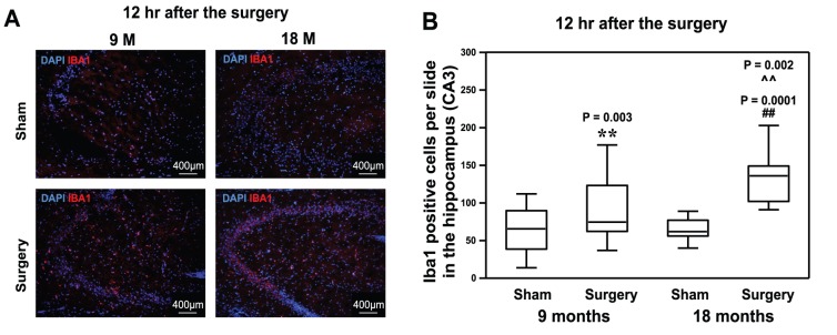
Figure 3. Peripheral surgical wounding increases the levels of Iba1 positive cells in the mouse hippocampus.

A. The immunohistochemistry staining showed that peripheral surgical wounding increases the levels of Iba1 positive cells in the hippocampus of both 9 and 18 month-old mice at 12 hours after the peripheral surgical wounding as compared to sham. There are more Iba1 positive cells in the hippocampus of 18 month-old mice than that of 9 month-old mice following the peripheral surgical wounding. B. The quantification of the images shows that the peripheral surgical wounding increases the number of Ibal positive cells in the hippocampus of both 9 and 18 month-old mice at 12 hours after the peripheral surgical wounding as compared to sham. Age potentiates the peripheral surgical wounding-induced increases in the levels of Iba1 cells. (** or ##: the difference between sham and peripheral surgical wounding group; ∧∧: the interaction between the group and the treatment). Iba1, ionized calcium-binding adapter molecule 1. N = 6.
