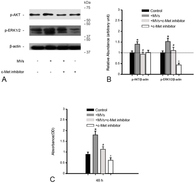
Figure 6. c-Met inhibitor abrogates the activation of AKT and ERK1/2 signaling in 786-0 cells and cell growth induced by MVs.
(A)(B) Gel photograph and densitometric analysis of p-AKT and p-ERK1/2 protein expression in 786-0 cells. MVs led to marked phosphorylation of AKT and ERK1/2 proteins after 48 h incubation with 786-0 cells, abolished by c-Met inhibitor. The density of each band was determined. Values in the graph are expressed as densitometry ratios of p-AKT/β-actin, p-ERK1/2/β-actin as folds over control (dotted line). *P<0.05, MVs vs. control; # P<0.05, MVs+c-Met inhibitor vs. MVs; × P<0.05, c-Met inhibitor vs. control; (C) CCK-8 Assay. After 48 h incubation with 786-0 cells, the addition of c-Met inhibitor greatly retarded cell growth induced by MVs. *P<0.01, MVs vs. control; # P<0.05, MVs+c-Met inhibitor vs. MVs; × P<0.05, c-Met inhibitor vs. control.

