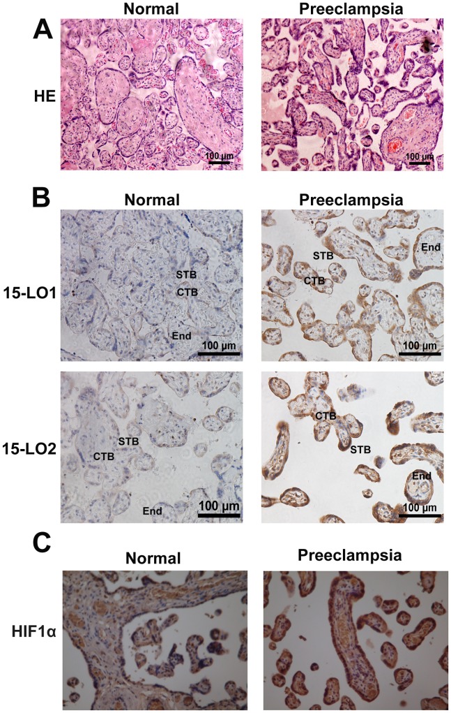
Figure 1. Morphologic changes in the placentas of normal and preeclamptic patients.
A. Hematoxylin–eosin (HE) staining. CTB: cytotrophoblast, which forms the inner layer of the trophoblast; STB: syncytrophoblast, which forms the surface of the villi; End: endothelial cell, a flat cell in the villi. Localization of 15-LO-1/2 (B) and HIF-1α (C). Increased expression of 15-LO-1/2 and HIF-1α gave rise to thicker syncytiotrophoblast membranes, more syncytial knots, and thicker vessels in preeclamptic placentas than in normal placentas (n = 10).

