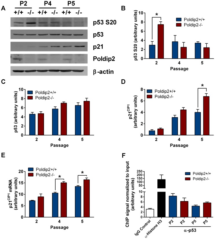
Figure 5. Poldip2 inhibits the p53 pathway.
(A) Immunoblotting was performed using lysates from Poldip2+/+ and Poldip2−/− MEFs in passages 2, 4 and 5. The blots were probed with antibodies against β-actin, Poldip2, p53, phospho-p53(S20), and p21CIP1. Densitometry was performed and corrected to β-actin (B, C, E). (D) p21CIP1 mRNA levels were assessed by RT-qPCR and corrected for the housekeeping gene PPIA. (F) ChIP was performed on Poldip2+/+ (blue) and Poldip2−/− (red) MEFs using p53 antibody and p21CIP1 promoter primers. Poldip2+/+ cells were used for the IgG negative and Histone H3 antibody positive controls. All samples were normalized to input DNA. Error bars represent mean ± SEM of 3–4 independent experiments. * P<0.05 comparing Poldip2+/+ with Poldip2−/−.

