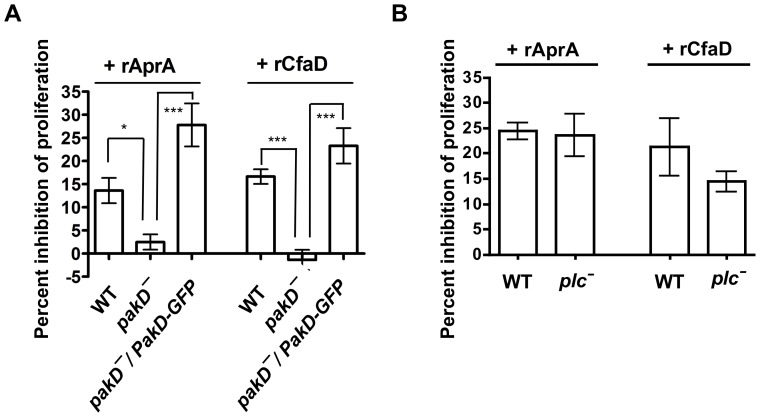
Figure 3. pakD– cells are insensitive to proliferation inhibition by AprA and CfaD.
(A) Log-phase cells were collected, resuspended to 0.5×10⁶ cells/ml, and either 300 ng/ml rAprA, 150 ng/ml rCfaD, or an equivalent volume of buffer was added to the cell culture. The inhibition of proliferation after 16 hours as a percent of the buffer control is plotted (100– ((experimental cell density/control cell density) * 100%)). “*” indicates that the difference in values between the conditions are significant with p<0.05, and “***” indicates p<0.001 (One-way ANOVA, Tukey's test). Values are mean ± SEM, n≥3. For both rAprA and rCfaD, the values for pakD– cells are not significantly different from a value of zero (p>0.05, paired t-test). For rAprA, but not for rCfaD, the differences between WT and pakD–/PakD-GFP are significant (p<0.05, One-way ANOVA, Tukey's test). (B) Wild-type and plc– cells were assayed for sensitivity to the proliferation-inhibiting activity of AprA and CfaD as described above. Differences between wild-type and plc– values are not significant (p>0.05, t-test).

