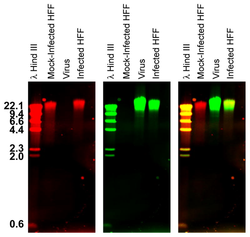
Figure 1. Dual color Southern blot.

DNA was extracted from uninfected or HCMV-infected human fibroblasts (HFF) as well as isolated HCMV viral particles. DNAs were separated on a 0.8% agarose gel before transferring to a charged nylon membrane. Subsequently the blot was hybridized with a mixture of Dig-labeled host genomic probes and biotin-labeled virus genomic probes. Probes were detected with IRDye 700-conjugated α-mouse secondary Ab and IRDye 800-conjugated streptavidin, respectively. The host and virus genomes were visualized simultaneously in the 700 (red- left panel) and 800 (green- middle panel) nm channels, respectively, on a Li-Cor Odyssey Infrared Imager. An overlay image of the red and green channels is shown at the right. The yellow color in the overlay image indicates lanes that contain both host and viral DNA. This figure has been modified from figure 7 in O’Dowd et. al. (O'Dowd et al., 2012).
