Fig. 2.
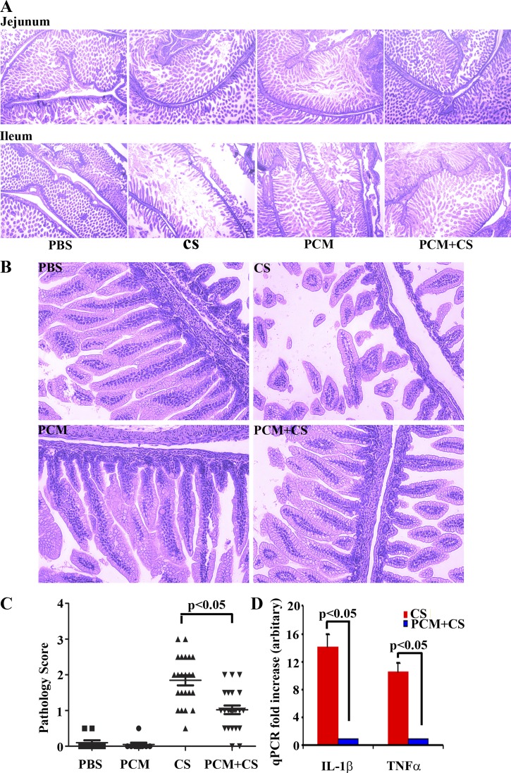
PCM treatment maintains the structural integrity of C. sakazaii-infected ileum. A and B: whole gut embedded in paraffin and processed by hematoxylin and eosin staining. The treatment condition is shown at the bottom of each column (×40 magnification; ×200 magnification) and at top left of each image. Villi displaying epithelial discontinuity, irregular alignment, and/or loss of epithelial cells was observed in the CS-infected group. In contrast, treatment with PCM and CS shows modest villus rupture only, which manifest regular alignment and no apparent loss of epithelial cells. Top, jejunal sections; bottom, ileal sections; ×40 magnification (A), ×200 magnification, ileal section (B). The pathology score (C) was determined according to the extent of epithelial sloughing, submucosal edema, neutrophil infiltration, and villus architecture destruction in the worst affected areas. Mean ± SE pathology score was graphed for 23–26 mice in each group and 2 sections were analyzed in each mouse (1-way ANOVA; post hoc Newman-Keuls multiple-comparison test: P < 0.05). D: real-time quantitative RT-PCR (qPCR) was performed in duplicate to measure the mRNA expression levels of the inflammatory cytokines IL-1β and TNF-α with GAPDH as an internal control. Mean ± SE fold increase was graphed (n = 10 mice in each group, Student's t-test: P < 0.05).

