
Fig. 4.

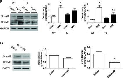
Increasing PKG activity reduced transforming growth factor (TGF)-β/Smad pathway in a UUO mouse model. A–D: kidney TGF-β1 mRNA and protein levels from UUO or sham group of mice were determined by real-time PCR and Western blotting, respectively. E: kidney sections from mice were stained with anti-TGF-β1 antibody. The positive staining is shown as brown. Representative light micrographs are shown. F and G: pSmad2 protein levels in kidney cortex from UUO or sham group of mice were determined by Western blotting. Data are presented as means ± SE (n = 5–6 mice/group). *P < 0.05 vs. WT/sham group or saline group. #P < 0.05 vs. WT/UUO group. &P < 0.05 vs. Tg/sham group.