
Fig. 7.
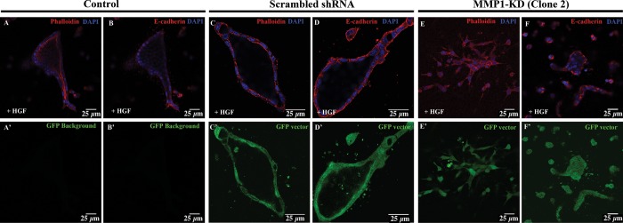
MMP1 KD disrupts tubule formation in 3D MDCK cells. 3D-cultured control (A and B), scrambled shRNA (C and D), and MMP1-KD (E and F) MDCK cells stained for phalloidin (red; A, C, and E) and E-cadherin (red; B, D, and F) are shown at 72 h post-treatment with HGF. At this time, control and scrambled shRNA cells are able to form polarized tubules with lumens (A–D). Control cells do not express GFP, whereas scrambled shRNA and MMP1-KD cells can be readily visualized using GFP coexpression as a marker (C′–F′). MMP1-KD cells fail to form polarized, lumen-filled tubules at 72 h after HGF treatment (E and F). Note that the distribution of actin is concentrated along the cell junctions in the MMP1-KD cells (E), in contrast with the enriched cortical actin expression, visible in the control cells (A). Scale bars = 25 μm.