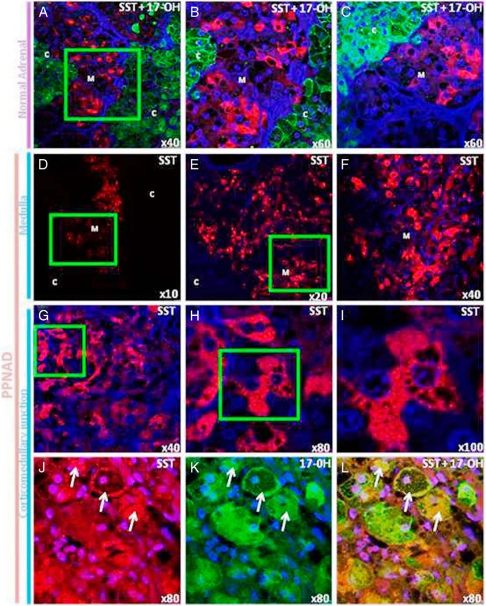
Figure 4.
Immunohistochemical localization of SST in normal adrenal gland and PPNAD tissues. A–C, Confocal laser scanning microscope photomicrographs showing the distribution of SST and 17α-hydroxylase immunoreactivities in the normal adrenal gland. The SST and 17α-hydroxylase immunoreactivities were revealed with Alexa 488-conjugated antibodies (red) and Alexa 594-conjugated antibodies (green), respectively. A, Anti-17α-hydroxylase antibodies produced diffuse labeling of the cortex, whereas SST-like immunoreactivity was exclusively detected in the medulla. B, High-magnification view of the field delimited by the green square in A. Medullary SST-positive cells presented as isolated cells or arranged in small clusters. C, High-magnification view of the corticomedullary junction showing that no SST immunoreactive cells could be observed in the inner cortex. D–I, Dual-channel confocal laser-scanning microscope analysis of the distribution of SST in PPNAD tissues. D–F, Different views at increasing magnifications showing diffuse SST labeling of the medulla in the adrenal gland removed from patient P1. G–I, Serial microphotographs at increasing magnification showing that SST labeling was also observed in groups of adrenocortical cells close to the corticomedullary junction containing numerous unstained lipid droplets (patient P2). J–L, Confocal laser-scanning microscope analysis of the corticomedullary junction in the PPNAD tissue removed from patient P5. J, Group of SST-positive cells (arrows) in the inner cortex close to the corticomedullary junction. K, The same SST-positive cells were also labeled by antibodies to 17α-hydroxylase. L, Merged microphotograph. C, cortex; M, medulla.

