
Figure 1.
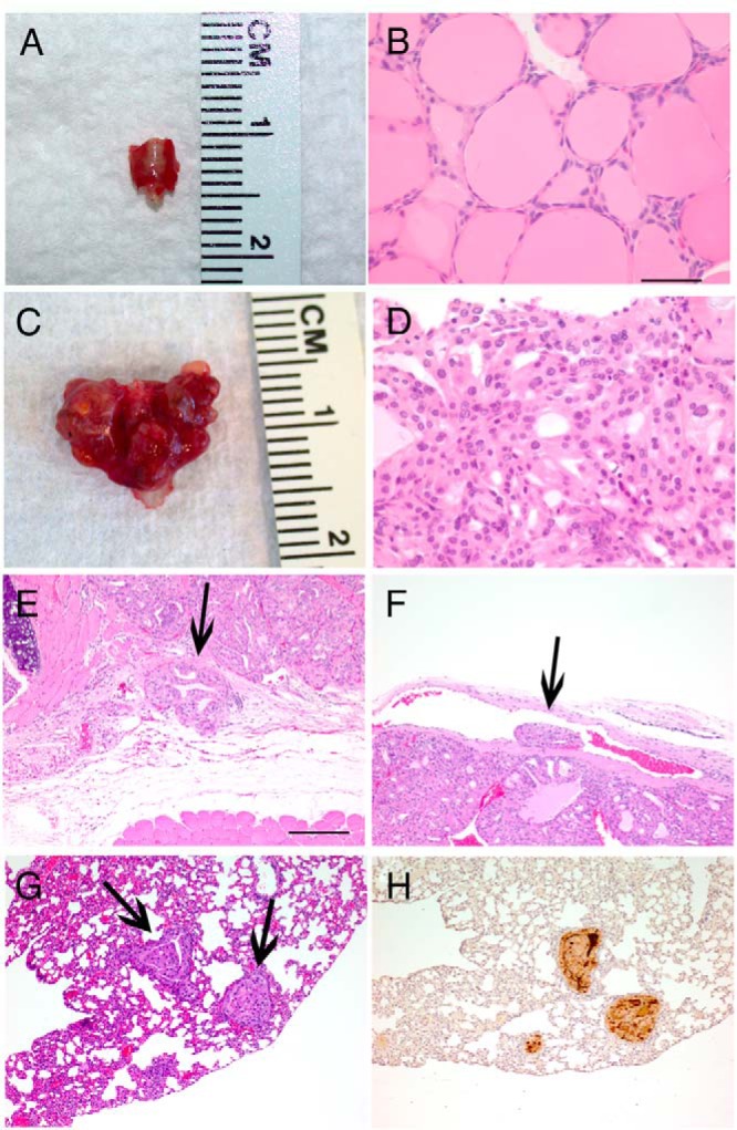
DRP-TpoKO tumors show consistent features of aggressive FTC and develop FTC-derived lung metastases. Macroscopic images of representative WT (A) and DRP-TpoKO (C) thyroids. Representative hematoxylin and eosin staining of high-magnification images of WT (B) and DRP-TpoKO (D) thyroids. Evidence of capsular (E, arrow) and vascular (F, arrow) invasion in DRP-TpoKO tumors. Representative photomicrographs of DRP-TpoKO follicular thyroid carcinoma lung metastases (arrows) stained with hematoxylin and eosin (F) and thyroglobulin (H). Scale bar (B, applies to D), 125 μm, (E, applies to F, G, and H), 500 μm.