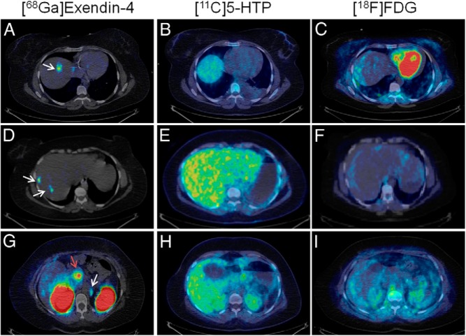
Figure 1.
[68Ga]Exendin-4 PET (left panels) confirmed several GLP-1R-positive lesions (white arrows) in the liver (A, D) and a para-aortal lymph node (G). No pancreatic or hepatic lesions could be conclusively detected by PET/CT using established tumor markers [11C]5-HTP (middle panels) and [18F]FDG (right panels). Each row shows the same transaxial abdominal sections for [68Ga]Exendin-4, [11C]5-HTP, and [18F]FDG, clearly demonstrating focal GLP-1R-positive lesions that could not be localized by established PET techniques. β-Cells in normal pancreas (red arrow) have significant expression of GLP-1R and can also be visualized by this technique (G). All images are normalized to a standardized uptake value of 1 with no background subtracted.

