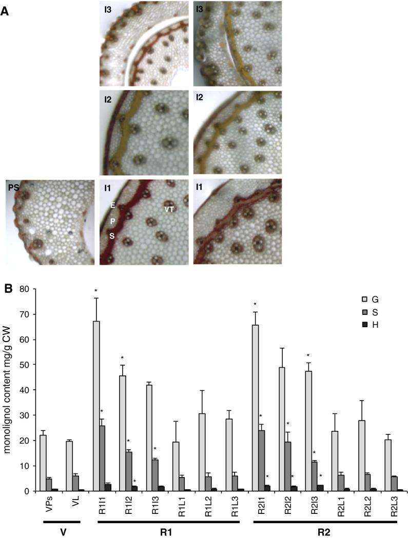
Fig. 4.
Composition of lignin in P. dilatatum stems and leaf blades through development as determined by a Mäule staining of transverse sections and b thioacidolysis of cell wall extracts. Asterisks indicate a significant difference (t test) relative to V stage pseudostems (P < 0.05). V Vegetative stage (Ps Pseudostem, L leaf), R1 early reproductive stage, R2 late reproductive stage (I1 internode 1, I2 internode 2, I3 internode 3, L1 leaf 1, L2 leaf 2, L3 leaf 3). Epidermal cells (E), Parenchyma cells (P), Sclerenchyma cells (S), Vascular tissue (VT), scale bar = 100 μm. mg/g CW = mg per gram of dry cell wall residues (mean and standard error of three biological replicates) G guaiacyl lignin, H hydroxyphenyl lignin, S syringyl lignin

