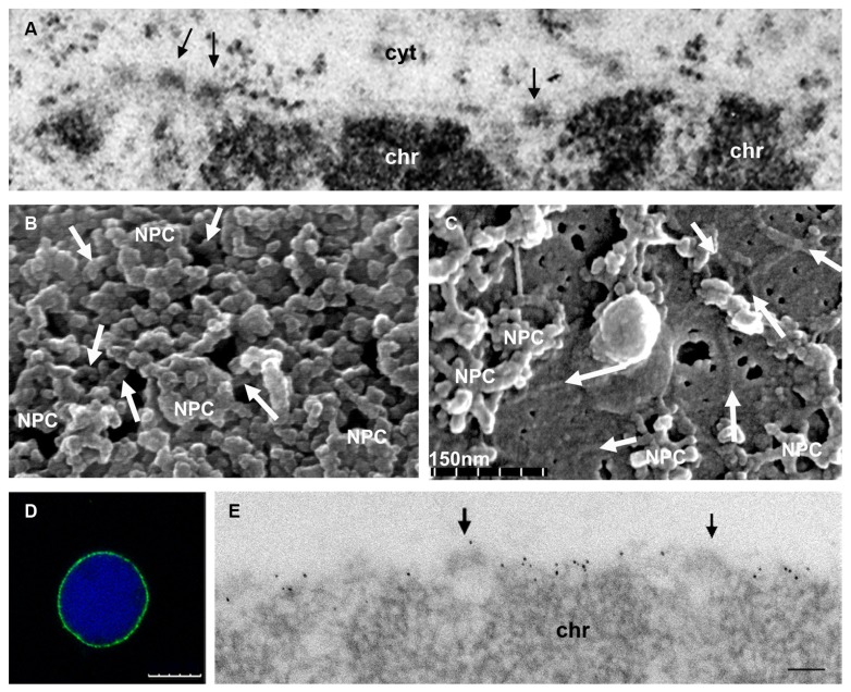
FIGURE 2.
Ultrastructure of the plant nuclear lamina and localization of the NMCP proteins. (A) Conventional thin section TEM image of the nuclear periphery of an onion meristematic root cell, showing a portion of the NE with its two membranes, and the dense NPCs (arrows) that traverse it. The heterochromatin (chr) is tightly attached to the INM but the thin lamina is not conspicuous with this technique. Cytoplasm (cyt). (B) Cytoplasmic face of the NE of a tobacco BY-2 cell nucleus extracted with Triton X-100 to remove the membranes and visualized by feSEM. The filaments of the lamina interconnecting the NPCs are evident (arrows). (C) feSEM image of the nucleoplasmic face of the NE of a BY-2 nucleus that has been fractured but not extracted with Triton X-100. Arrows indicate the filaments of the lamina in the membrane. (D,E) Detection of NMCP1 in the lamina of isolated meristematic onion nuclei extracted with Triton X-100 after immunofluorescence and DAPI staining (D), or TEM immunogold labeling (E). After removing the membrane, the lamina with a lower electron density than chromatin and containing NMCP1 proteins is evident at the nuclear periphery. The association with NPCs (arrows) and the tight attachment to the condensed chromatin masses (chr) can be seen. (B,C courtesy of Drs J. Fiserova and M. W. Goldberg). Bars in D = 10 μm and in E = 100 nm.

