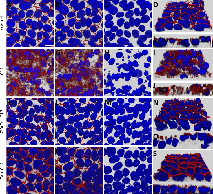
Fig. 2.
C12 causes reorganization/disassembly of tight junctions [zonula occludens 1 (ZO-1)] in Calu-3 epithelial cells, and ZVAD and Tg prevent this response. Calu-3 cell monolayers grown on Snapwell inserts and mounted in Ussing chambers for measurement of transepithelial parameters were fixed and labeled with an anti-ZO-1 antibody (red) and stained for nuclei (Hoechst 33342, blue). A–E: untreated control epithelium. x-y images of ZO-1 and nuclei were viewed from the apical side (A) and basolateral side (B), and nuclei alone were viewed from the basolateral side (C). Angled-view projection of ZO-1 and nuclei from the basal surface of a confocal image stack is shown in D; selected orthogonal cut (x-z) view with apical side pointing upward is shown in E. ZO-1 label was prominent at the perimeters of the cells, and there was no apparent ZO-1 staining on the basolateral side. Nuclei were oblong or oval. F–J: C12-treated monolayer; image projections as described in A–E. ZO-1 strands appeared to be disassembled, with distribution throughout the cell and appearance on lateral and basal sides of some cells. Nuclei generally were shrunken, and many appeared to have fragmented. K–O: ZVAD + C12-treated culture from recording in Fig. 1A; image projections as described in A–D. ZO-1 appeared normal, with apical strands surrounding large, oval nuclei. P–T: Tg + C12-treated culture from recording in Fig. 1B; image projections as described in A–E. ZO-1 appeared normal with apical strands; nuclei were large and oval. Scale bar, 10 μm. Each image is typical of images from 3 experiments.

