Fig. 3.
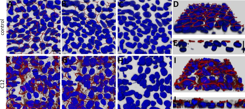
C12 causes reorganization/degradation of tight junctions in primary cultures of airway epithelia. Well-differentiated primary airway epithelial cell cultures grown on filters at an air-liquid interface were fixed, labeled with an anti-ZO-1 antibody (red), and stained for nuclei (Hoechst 33342, blue). A–E: untreated control epithelium. x-y images of ZO-1 and nuclei were viewed from the apical side (A) and basolateral side (B), and nuclei alone were viewed from the basal side (C). Angled-view projection of ZO-1 and nuclei from the apical surface of a confocal image stack are shown in D; selected orthogonal cut (x-z) view with apical side pointing upward is shown in E. ZO-1 label was prominent at the perimeters of the cells. Nuclei were oval or oblong. F–J: monolayer treated for 3 h with 100 μM C12; image projections as described in A–E. ZO-1 strands appeared to be disassembled in large patches of cells, with distribution throughout the cell and appearance on lateral and basal sides of some cells. Nuclei appeared misshapen in many cells. Scale bar, 10 μm. Each image is typical of images from 2 experiments.

