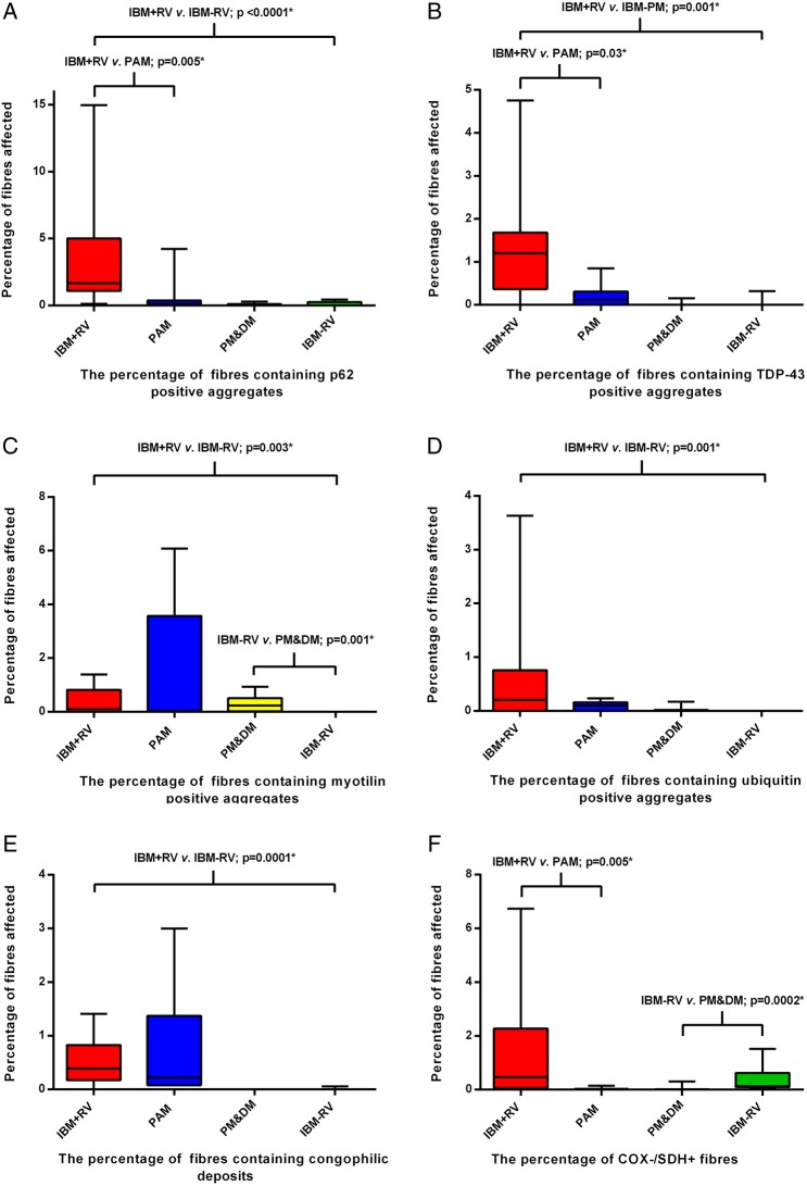
Figure 3 .
Box and whisker plots illustrating the percentage of fibres in each diagnostic category containing p62 (A), TDP-43 (B), myotilin (C) and ubiquitin (D) immunoreactive aggregates, congophilic deposits (E) and COX−/SDH+ fibres (F). All protein aggregates were present in a greater percentage of fibres in IBM+RV than in IBM - RV. There was no difference in the percentage of COX−/SDH+ muscle fibres between these groups. IBM+RV biopsies had a greater percentage of fibres containing p62 (A) and TDP-43 (B) immunoreactive aggregates and COX−/SDH+ fibres (F) than PAM. Pathological findings were similar in IBM - RV and PM&DM, with no differences in the percentage of fibres containing p62 (A), TDP-43 (B) and ubiquitin (D) immunoreactive aggregates or congophilic deposits (E). However, there was a greater percentage of COX−/SDH+ fibres (F) in IBM - RV than PM&DM and a greater percentage of fibres containing myotilin immunoreactive aggregates (C) in PM&DM than IBM - RV. *Statistically significant results. COX, cytochrome oxidase; IBM, inclusion body myositis; PAM, protein accumulation myopathies with rimmed vacuoles; PM&DM, steroid-responsive inflammatory myopathies; SDH, succinate dehydrogenase; TDP-43, transactivation response DNA binding protein-43.

