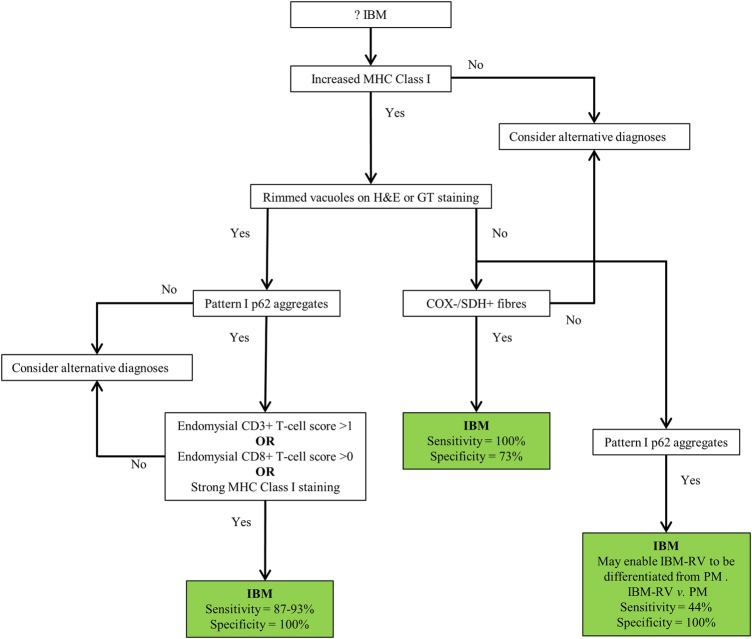
Figure 4 .
Proposed diagnostic algorithm for IBM based on pathological findings. Increased MHC class I staining was observed in all cases of IBM and pattern I p62 aggregates in all cases of IBM+RV making them good initial screening tests. Their absence rules out a diagnosis of IBM and IBM+RV, respectively. The presence of endomysial CD3 T-cell score >1, endomysial CD8 T-cell score >0 or strong MHC class I staining in a biopsy with rimmed vacuoles and pattern I p62 aggregates secures a diagnosis of IBM+RV. Differentiating IBM - RV and PM&DM pathologically is more challenging. The presence of COX−/SDH+ fibres is not specific to IBM - RV; although COX−/SDH+ fibres were not present in every case of IBM - RV their absence casts doubt on the diagnosis of IBM - RV. Pattern I p62 aggregates may enable IBM - RV to be differentiated from PM when present. However, they may lack sensitivity for IBM - RV and therefore, their absence does not rule out the diagnosis. COX, cytochrome oxidase; GT, Gomori trichrome; IBM, inclusion body myositis; MHC, major histocompatibility complex; PM&DM, steroid-responsive inflammatory myopathies; SDH, succinate dehydrogenase.

