Figure 6.
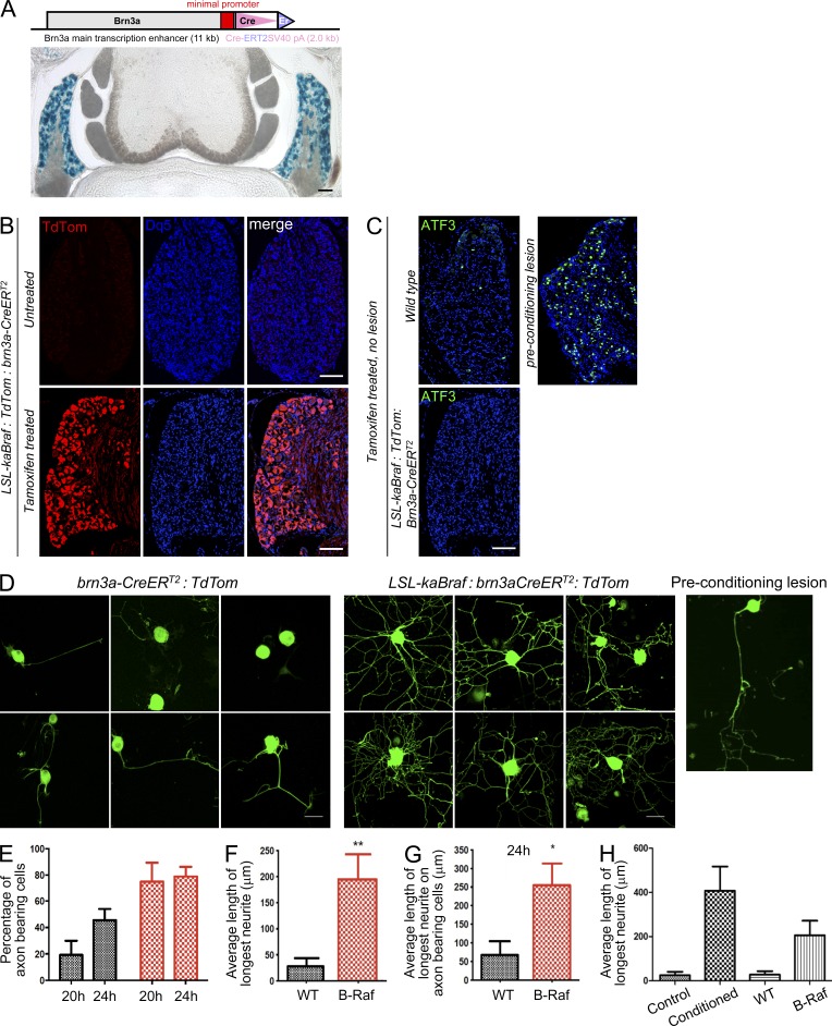
Activation of B-RAF signaling in mature DRG neurons elevates their growth competency. (A, top) Schematic of the brn3a-CreERT2 construct used to generate the brn3a-CreERT2 deleter mouse line. (bottom) A cross section of the spinal cord of a 10-wk-old Rosa26-lacZ:brn3aCreERT2 mouse treated with tamoxifen. Blue LacZ staining indicates CreERT2-medicated recombination in the DRG neurons. (B) Representative DRGs from adult LSL-kaBraf:TdTom:brn3a-CreERT2 mice without (top left) and with (bottom left) tamoxifen treatment. TdTom expression indicates recombination in DRG neurons. Cells were counterstained with Draq5 (Dq5) to label nuclei. (C) ATF3 is induced by preconditioning lesion. Blue shows nuclear stain Draq5. (D) Representative images of adult DRG neurons derived from intact brn3a-CreERT2:TdTom (left), LSL-kaBraf:brn3aCreERT2:TdTom (middle), and WT preconditioning lesioned mice (right) after 24 h in vitro. TdTom is shown in green to improve contrast. Bars: (A–C) 100 µm; (D) 20 µm. (E–H) Quantitation of axon extension in adult DRG cultures at 24 h in vitro. Data were collected from three independent experiments from three animals per genotype or condition and analyzed as described previously (Parikh et al., 2011); >100 cells were counted per group. Error bars indicate SEM. One-way ANOVA with post-hoc Tukey’s HSD test: *, P < 0.01; **, P < 0.005.

