Figure 8.
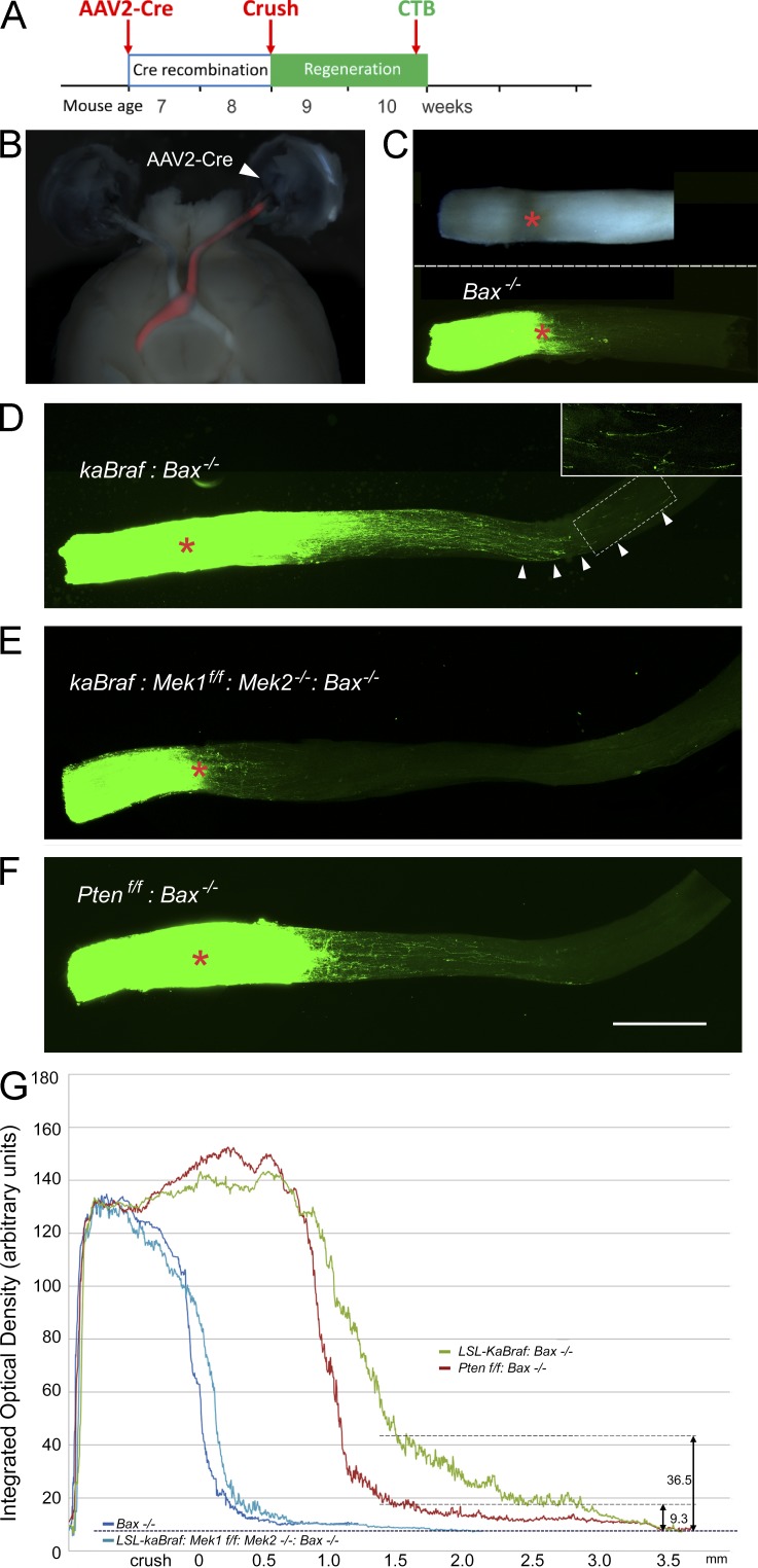
Activation of B-RAF enables regenerative axon growth in the crush-lesioned optic nerve via the canonical effectors MEK1/2. (A) Schematic of experimental time course. (B) Intravitreal injection of AAV2-Cre induces expression of TdTom in retinal ganglion neurons, labeling the entire optic nerve (red). (C, top) Whole-mount image of a crushed Bax−/− optic nerve. Crush site is indicated by a red asterisk here and in all following panels. (bottom) Confocal fluorescence image of the same nerve. Green shows axons anterogradely labeled with CTB–Alexa Fluor 488. (D) Whole-mount confocal imaging shows strong regenerative growth in the lesioned kaB-RAF–expressing optic nerve. (inset) Axons at ∼3.5 mm from the crush site (magnified from the boxed area). Arrowheads indicate outgrowing axons. (E) Loss of MEK1 and MEK2 abolishes the regeneration driven by kaB-RAF. (F) Optic nerve regeneration in the absence of PTEN. Bar, 0.5 mm. (C–F) Images are representative of three optic nerves per genotype. (G) Quantitation of axon regenerative growth in the optic nerve 2 wk after nerve crush; genotypes as shown in B–E. At ∼1.6 mm from the crush site, the density of regenerating axons is more than threefold greater in the LSL-kaBraf:Bax−/− genotype than in the Ptenf/f:Bax−/− genotype. Data are from three nerves per genotype. Optic densities were acquired from the whole-mount optic nerves using an LSM710NLO two-photon confocal microscope with the ZEN2009 software. Data were normalized by setting the baseline OD, as measured 0.2 cm proximal to the crush site in all nerves, to the same (arbitrary) level.

