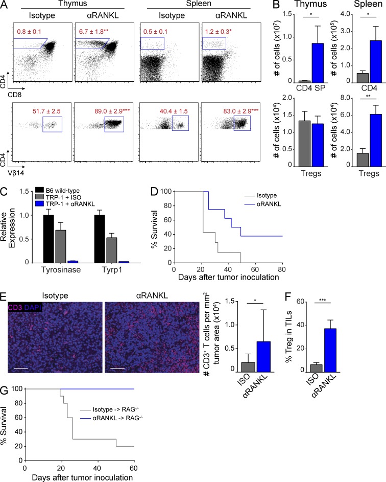
Figure 5.
RANKL blockade increases anti-melanoma immune response. (A and B) RAG1−/− x TRP-1 TCR Tg mice were treated with isotype or anti-RANKL antibody for 2 wk and thymus and spleen were harvested for analysis. Flow cytometry plots in A show percentage of CD4+ (top) and CD4-gated Vβ14+ (bottom) T cells. Bar graphs in B show quantification of data in A. Values represent mean ± SEM. *, P ≤ 0.05; **, P ≤ 0.01; ***, P ≤ 0.001, Student’s t test. Data shown is representative of at least three independent experiments with n = 4–7 mice per group. (C) Ear skin from mice treated with isotype (gray) or anti-RANKL (blue) antibody was analyzed by qPCR for Tyrosinase and Tyrp1 expression. Results standardized to β2m and normalized to untreated B6 wild-type mice (black) with bars depicting mean ± SD. (D) Survival curves of RAG1−/− x TRP-1 TCR Tg mice inoculated with B16 melanoma after treatment with isotype (gray) or anti-RANKL (blue) antibody, n = 7–8 mice for each group. P ≤ 0.05, Log-rank test. Data shown is representative of four independent experiments. (E) Tumor sections from isotype or anti-RANKL–treated mice in D were immunostained for CD3 (pink) with DAPI counterstain (purple). Bars, 50 µm. Mean densities of CD3+ cells per tumor area are quantified on the right. Four sections were evaluated for each treatment group and 6 imaging fields were randomly scored from each section. Graphs depict mean ± SEM. *, P ≤ 0.05, Student’s t test. (F) RAG1−/− x TRP-1 TCR Tg mice were inoculated with B16 melanoma after treatment with isotype (gray) or anti-RANKL (blue) antibody. Mice were harvested at 21 d after tumor inoculation and tumor-infiltrating lymphocytes (TILs) were analyzed by flow cytometry. Graph depicts percentage of Foxp3+ CD25+ T reg cells among CD4+ T cells. Values represent mean ± SEM. ***, P ≤ 0.001, Student’s t test. Data shown is n = 7–9 mice per group pooled from two independent experiments. (G) Survival curves of RAG1−/− recipients inoculated with B16 melanoma after adoptive transfer of splenocytes from either anti-RANKL (blue)– or isotype (gray)–treated RAG1−/− x TRP-1 TCR Tg mice, n = 10 mice for each group. P ≤ 0.001, Log-rank test. Data shown is representative of two independent experiments.

