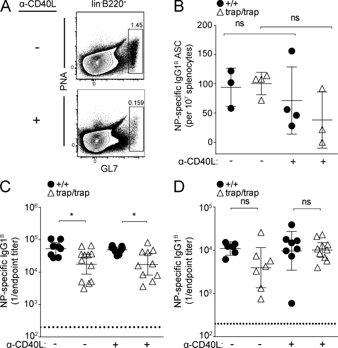
Figure 6.
ZBTB20 deficiency causes plasma cell–intrinsic defects. (A) Flow cytometric quantification of germinal center B cells after administration of blocking α-CD40L antibody. Wild-type mice were immunized with alum-adjuvanted NP-CGG and then left untreated or injected with 200 µg α-CD40L at days 13, 14, and 15. Frequencies of splenic lineage− (CD4/8/Gr-1/Ter119) germinal center B cells were analyzed as shown at day 16 after immunization. Data are representative of two experiments with two mice per treatment group. (B) ELISPOT analysis of splenic plasma cells at 4 wk after immunization with TLR ligand–adjuvanted NP-CGG. Mice were left untreated or administered α-CD40L at days 13, 14, and 15 as in A. Statistical significance was determined with an unpaired Student’s two-tailed t test. Error bars depict mean values ± SD. Each data point represents one mouse. Data are representative of two experiments, each with three to four mice per group. (C and D) ELISA measurements of serum antibody titers (error bars depict geometric means ± 95% confidence interval) from Zbtb20+/+ and Zbtb20trap/trap fetal liver chimeras immunized with alum (C)- or TLR ligand–adjuvanted (D) NP-CGG and then treated with α-CD40L or left untreated as in A. NP-specific serum antibody titers were quantified at 7 wk after immunization. Data are cumulative from two experiments with 8–12 (C) or 6–10 (D) mice per genotype. Statistical significance was determined with a Mann–Whitney test. ns, not significant (P > 0.05); *, 0.01 < P < 0.05.

