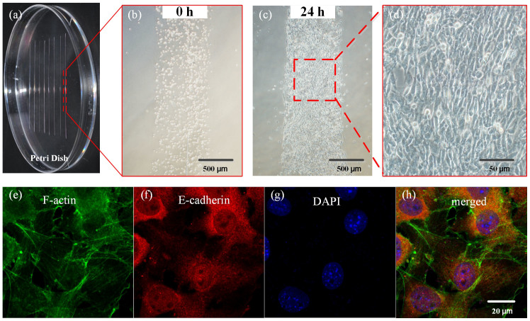
Figure 2. Point-of-care tissue engineering.
(a) Writing scar patch assays (3T3 fibroblasts and collagen hydrogel) on a Petri dish. Phase contrast images of cells in one scar patch immediately after writing (b) and after 24 hours of culture (c and d). Fluorescent immunostaining for F-actin (e), E-cadherin (f) and DAPI (g). The merged image (h) shows a mature cellular network with E-cadherin connections.

