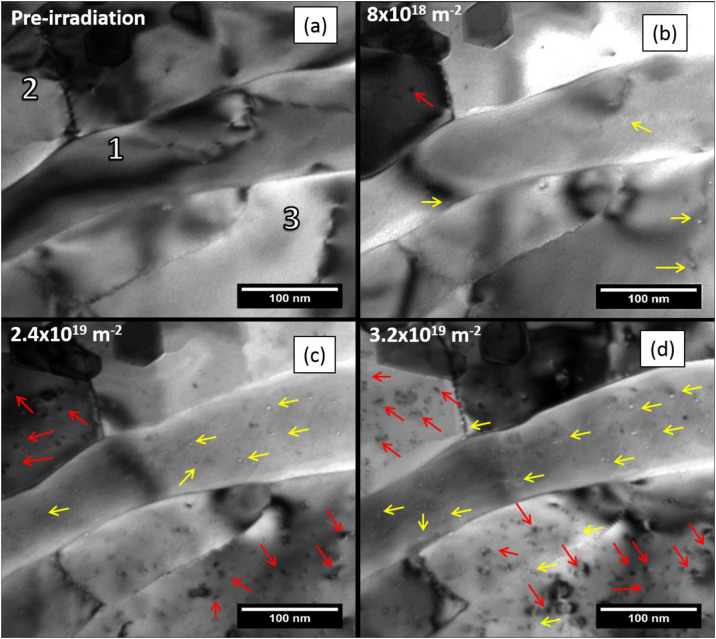
Figure 1.
TEM micrographs of in situ 2 keV He+ ion irradiation of tungsten at 950°C showing: (a) nanocrystalline (1) and ultrafine (2 and 3) grains before irradiation; (b) at a fluence of 8 × 10¹⁸ ions.m−2 and after bubble nucleation (bubbles indicated by yellow arrows); (c) after irradiation to a fluence of 2.4 × 10¹⁹ ions.m−2 showing point defect cluster formation (indicated by red arrows) occurred predominantly in grains 2 and 3; and (d) after irradiation to a fluence of 3.2 × 10¹⁹ ions.m−2 with a higher areal density of point defect clusters and small dislocation loops evident in grains 2 and 3 whilst grain 1 demonstrates a uniform distribution of bubbles and a significantly lower areal density of defect clusters and dislocation loops. (arrows guide the eye to aid in identifying respective defects).

