
FIG 1.
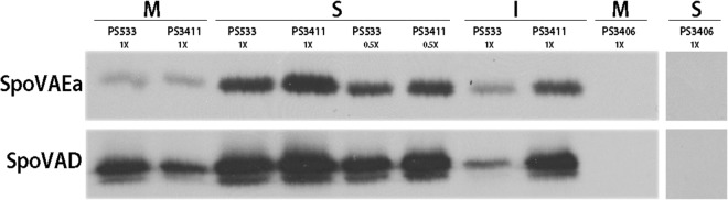
Detection of SpoVAEa and SpoVAD in spores and their distribution in different spore fractions. Spores of B. subtilis strains PS533 (wild type), PS3411 (↑spoVA mutant), and PS3406 (sleB spoVA mutant) were purified and decoated, removing some spore coat protein, as well as the spore outer membrane. The spores were then disrupted and fractionated as described in Materials and Methods, giving the spore IM fraction (M), the soluble fraction (S) consisting of soluble proteins from the spore core, and the integument fraction (I) consisting of insoluble coat protein, partially degraded spore cortex, and IM that is not sheared off the I fraction (9, 25–27). Aliquots of the various fractions from equal numbers of spores (∼108) were then subjected to Western blot analysis with antiserum against SpoVAEa and SpoVAD. All samples were run on the same Western blot, except the S fraction from PS3406 spores. The purified SpoVAD protein migrated identically to the SpoVAD antigen detected in spores, while the SpoVAEa antigen detected in spores ran more slowly than the His-tagged full-length SpoVAEa protein used as a marker (data not shown).